Figure 5.
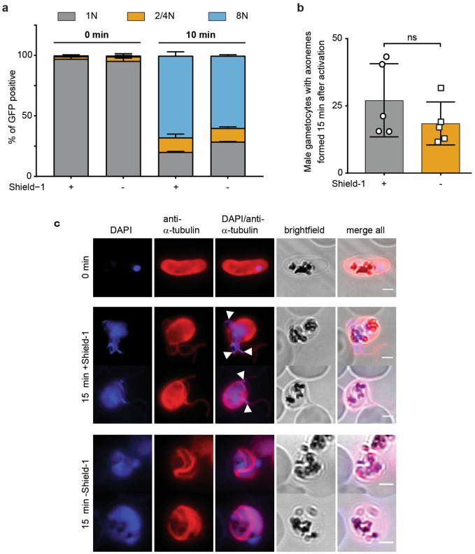
Detailed phenotyping of the exflagellation defect in NF54/MAP-2GFPDD parasites. (a) Ploidy of GFP-positive microgametocytes 0 and 10 min after activation of gametogenesis under protein-stabilizing (+ Shield-1) and -degrading (− Shield-1) conditions assessed by flow cytometry. Parasites were split (± Shield-1) 24 h before analysis. Values show the means ± SD of three biological replicates. (b) Number of male gametocytes that form axonemes 15 min after activation of gametogenesis, determined from anti-α-tubulin IFAs. Protein-stabilizing (+ Shield-1) and -degrading (− Shield-1) culturing conditions are compared. Parasites were split (± Shield-1) 24 h before analysis. Values show the means ± SD of five biological replicates with individual data points represented by open symbols. ns, not significant (paired two-tailed Student’s t test). (c) Representative images of anti-α-tubulin IFAs showing axoneme formation 15 min after activation of gametogenesis. Protein-stabilizing (+ Shield-1) and -degrading (− Shield-1) culturing conditions are compared. White arrowheads indicate incorporation of DNA into newly forming microgametes exclusively in parasites cultured under protein-stabilizing (+ Shield-1) conditions. Parasites were split (± Shield-1) 24 h before probing. Scale bar = 2 µm.

