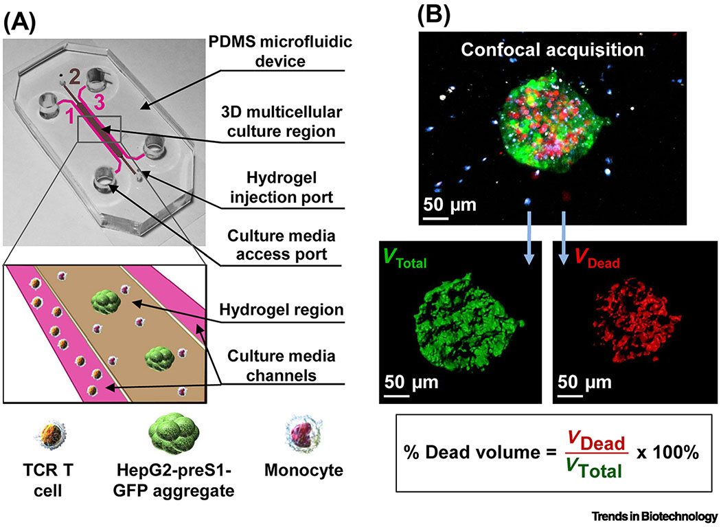
Figure 1. MPS modeling of the infiltration and cytotoxicity of therapeutic lymphocytes.
(A) To study the influence of monocytes on tumor cell killing by TCR-engineered T cells in the tumor microenvironment, a simple and customizable microfluidic device was developed for the 3D culture of HepG2 hepatocellular carcinoma tumor spheroids expressing the preS1 portion of the hepatitis B virus envelope protein linked to green fluorescent protein (preS1-GFP) and monocytes in a central collagen hydrogel. This was flanked by two media channels with or without TCR-engineered T cells specific for the hepatitis B virus antigen present on the tumor cells. (B) Tumor cell killing was assessed by confocal imaging of pre-labeled tumor spheroids (green) surrounded by monocytes (blue) and T cells (white). Dead tumor target cells were labeled by the live/dead cell exclusion dye DRAQ7 (red), and the volume of dead cells was quantified relative to the total spheroid volume. Reproduced from [16] under a Creative Commons license.

