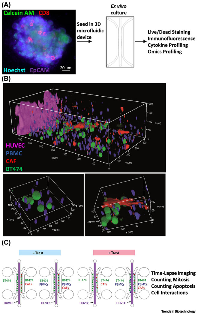
Figure 3. MPS modeling of complex cell types in the tumor immune microenvironment.
(A) In one approach, patient tumor organoids can be harvested to retain the autologous stromal cells and immune cells prior to embedding in 3D microfluidic hydrogel devices. Confocal imaging of a patient high-grade serous ovarian carcinoma organoid containing viable (calcein acetoxymethyl ester (AM), green), EpCAM+ epithelial cells (purple) demonstrated the presence of CD8+ T cells (red) and all nucleated cells (Hoechst, blue). Microfluidic devices have facilitated the identification of cytokine profiles predictive of differential sensitivity to PD-1 blockade and exogenous agents that enhance sensitivity. Adapted from [28] under a Creative Commons license. (B) To provide for tunable control over cell density, type, ratio, and orientation, a “tumor ecosystem-on-a-chip” was developed (top). Confocal imaging of one half of the device demonstrated the position of pre-labeled human BT474 breast cancer cells (green), PBMC including immune cells (blue), and human breast Hs578T CAF (red) parallel to a central HUVEC monolayer (magenta). Magnified 3D images show physical interactions between various cell types (bottom). (C) Experimental design of the microfluidic devices containing two hydrogel compartments (gray) containing the indicated cell type configurations to allow for the study of how ADCC stimulated by the therapeutic antibody trastuzumab (Trast) is antagonized by CAF. Adapted from [30] under a Creative Commons license.

