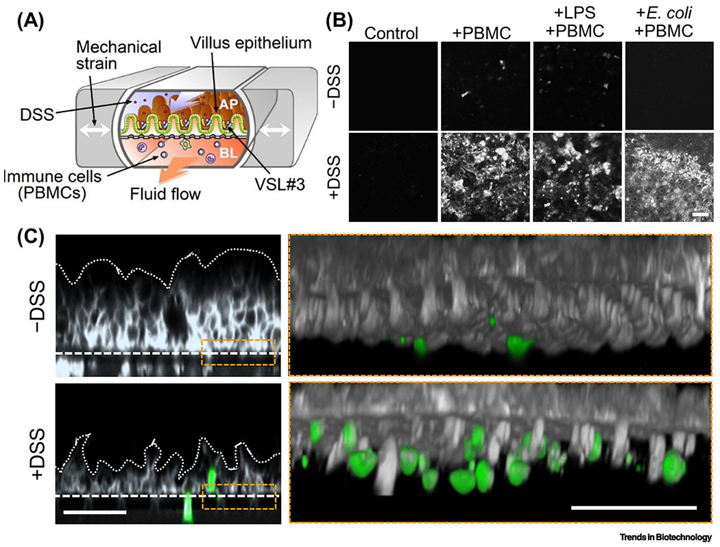
Figure 4. MPS modeling of intestinal inflammation.
(A) A schematic of the human gut inflammation-on-a-chip that provides the accessibility and versatility to manipulate the complex inflammatory milieu. Arrows in the central microchannels and the vacuum chambers indicate the directions of fluid flow and the peristalsis-like mechanical distortions, respectively. AP, apical; BL, basolateral. (B) Top-down views of the fluorescently labeled villous epithelium under the oxidative stress in the presence or absence of dextran sodium sulfate at various combinations of inflammatory triggers (PBMC, LPS, and E. coli) in the human gut inflammation-on-a-chip. Bar, 50 μm. (C) Cross-sectional views of the immune cell recruitment visualized in situ in the gut inflammation-on-a-chip. Gray, microengineered villous epithelium; green, PBMC. Dotted lines indicate the contour of the 3D villi. Dashed lines show the location of the basement membrane in the gut inflammation-on-a-chip. Bars, 50 μm. B and C reproduced from [60] with permission.

