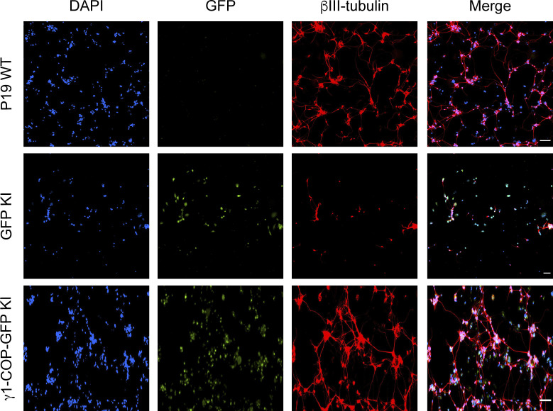
Figure S5. Inhibition of neurite outgrowth in P19 GFP knock-in (KI) cells.
Representative fluorescence microscopy images of P19 WT, GFP KI, and γ1-COP-GFP KI as indicated at day 8 of differentiation to analyze the expression of the neuronal marker βIII-tubulin (indirect immunofluorescence) and GFP (direct fluorescence). Scale bar is 100 μm.

