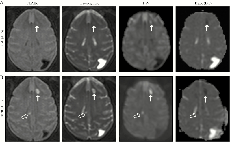
Figure 2.
(A), A small focal lesion (arrows) appeared on day 15 after exposure in the left frontal subcortical white matter in 1 animal (8078). (B), Progression of disease is noted on day 17 (terminal day), with a total of 7 abnormal signal intensities now seen bilaterally. Incidental note is made of a preexisting congenital abnormality in this animal (colpocephaly, dilatation of the left posterior occipital horn). Abbreviations: DW, diffusion weighted; FLAIR, fluid-attenuated inversion-recovery; trace (DT), trace of diffusion tensor.

