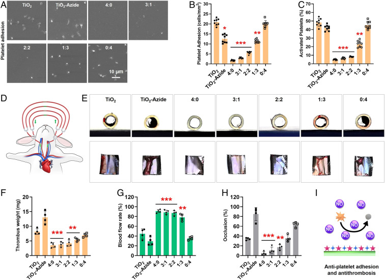
Fig. 3.
(A) SEM images, (B) average density, and (C) activation rates of the adhered platelets after incubation with different 316L SS substrates supplemented with NO donor. (D) Schematic illustration of the rabbit AV shunt model. (E) Cross-sectional photographs of tubing and the corresponding thrombus formed in different groups. Quantitative results of (F) the thrombus weight, (G) blood flow, and (H) occlusion rate in different groups. (I) Schematic antiplatelet adhesion and activation on the cografted surface. Statistically significant differences are indicated by *P < 0.05, **P < 0.005, ***P < 0.001 compared with the bare surface (the TiO2 group).

