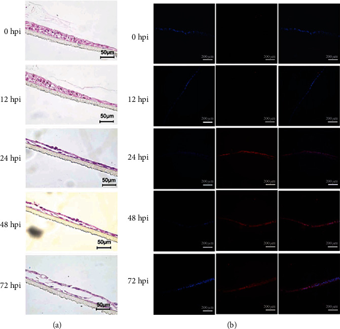
Figure 6.

H1N1 infection of 3D ALI cultures of HNTEC. (a) Morphology of 3D ALI cultures of HNTEC infected by H1N1pdm. HNTEC were cultured in 3D ALI as indicated in days and then inoculated with viruses at MOI of 0.01 at the apical layer. Virus inoculums were removed, after 1 hr adsorption. The cultures were rinsed with PBS for 3 times and replenished with fresh medium. At the indicated time points, 3D cultures were harvested. The 3D ALI cultures were then fixed, paraffin-embedded, and sectioned. Histology was captured under the microscope. Scale bar, 50 μm. (b) Expression of influenza A virus nucleoprotein in 3D ALI cultures after H1N1pdm infection. 3D ALI cultures with H1N1pdm were stained with the antibody against anti-influenza A virus nucleoprotein. Expression of influenza A virus nucleoprotein detected by immunofluorescence protocols. The nuclear staining was used with 0.5 μg/ml DAPI. Scale bar, 100 μm.
