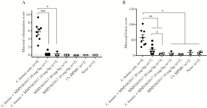
Figure 5.
Mucosal colonization and lesion scores in piglets. (A) Colonization scores; (B) lesion scores. Mucosal colonization was scored from 0 to 5 (0 = no infection detected; 1 = 1%–20% epithelial surface infected; 2 = 21%–40% surface infected; 3 = 41%–60% surface infected; 4 = 61%–80% surface infected; 5 = 81%–100% surface infected) in 3 fields from each anatomic site: duodenum, jejunum, ileum, cecum, and spiral colon and then averaged. For each piglet, the final score is a sum of the averages (A). Mucosal lesion scores were generated for each site for changes in villi, epithelial cells, and inflammation. The following scores were used: normal = 0, equivocal = 2.5, mild = 5, moderate = 10, marked = 15. The final lesion score was a sum of scores from duodenum, jejunum, ileum, cecum, and colon for each piglet. The scatter plot (A and B) represents mean ± standard error of the mean. A Mann-Whitney test was conducted using GraphPad Prism 7.03: *, P < .05; **, p<.01; ***, P < .001. Cryptosporidium hominis-infected group (n = 8, closed circle [●]); C hominis-infected and MMV665917 (20 mg/kg)-treated group (n = 7, closed square [■]); MMV665917 (20 mg/kg)-treated group (n = 4, open square [□]); C hominis-infected and MMV665917 (10 mg/kg)-treated group (n = 3, closed triangle [▲]; MMV665917 (10 mg/kg)-treated group (n = 2, open triangle [△]); 1% hydroxypropyl methyl-cellulose (HPMC)-treated group (n = 2, open rhombus [◊]); and nontreated control (n = 2, open circle [○]).

