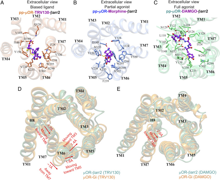
Fig. 7.
The pp-μOR binding pocket after ∼500 ns of MD simulation stabilized by the βarr2 in the presence of (A) TRV130, a biased agonist; (B) morphine, a partial agonist; and (C) DAMGO, a full agonist for the βarr2 coupling. The dotted lines represent the hydrogen binding. (D) The structural differences between the cytoplasmic region of the μOR after recruiting the Gi protein (orange) and the βarr2 (green) in the presence of TRV130. The red arrows represent movements of the μOR induced by the βarr2. (E) The structural differences between the cytoplasmic region of the μOR after recruiting the Gi protein (orange) and the βarr2 (green) in the presence of DAMGO. The red arrow represents the only significant movement of the μOR induced by the βarr2.

