Fig. 1.
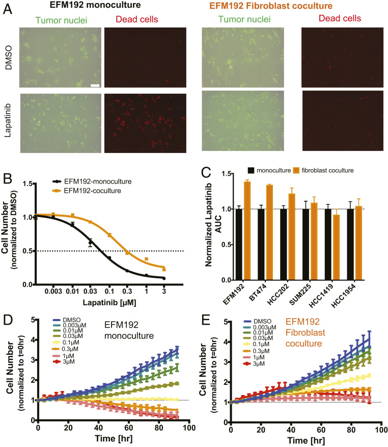
Fibroblasts limit lapatinib response in a subset of HER2+ breast cancer cell lines in vitro. (A) H2B-GFP (green) expressing EFM192 tumor cells cocultured with AR22 fibroblasts for 96 h under control (dimethyl sulfoxide [DMSO]) and lapatinib (1 μM) treatment. Representative images from three biological replicates of monoculture and coculture of viable (green only) and dead (orange) tumor cells (red objects: ethidium bromide staining). (Scale bar, 200 μm.) (B) Cells were incubated with increasing drug concentrations for 96 h and the number of tumor cells was assayed in monoculture (black) and AR22 coculture (orange). Data are representative of three independent experiments and error bars are SD for three replicate wells. (C) Lapatinib AUC values. Data are derived from three independent experiments and error bars are SEM for three biological replicates. (D and E) Change in viable EFM192 tumor cell numbers over time at increasing lapatinib concentrations in monoculture and coculture with AR22 fibroblasts. Data are representative of three independent experiments and error bars correspond to SD for n = 3 replicate wells.

