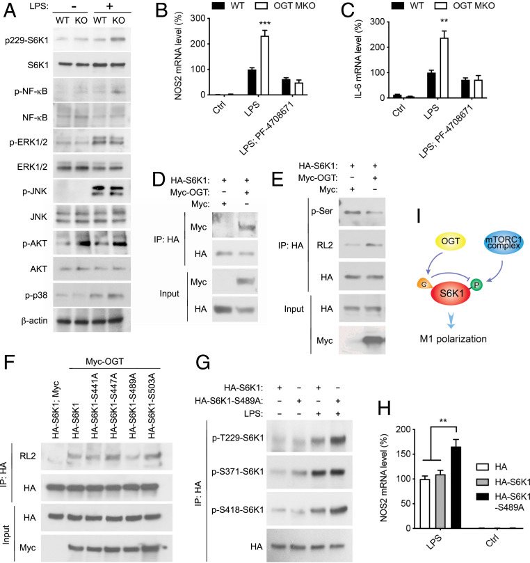
Fig. 7.
OGT inhibits macrophage proinflammatory polarization by suppressing mTORC1/S6K1 signaling. (A) Western blot analysis showing the activation of S6K1, NF-κB, ERK, JNK, p38 MAPK, and Akt in unstimulated and LPS-stimulated (30 min) WT and OGT KO peritoneal macrophages. (B and C) Nos2 and Il-6 mRNA levels in unstimulated, LPS-stimulated, and PF-04708671-pretreated LPS-stimulated WT and OGT KO BMDMs (n = 4). (D) Immunoprecipitation (IP) and Western blot analysis showing the interaction between exogenously expressed HA-S6K1 and Myc-OGT in HeLa cells. (E) IP and Western blot analysis showing that OGT overexpression enhances S6K1 O-GlcNAcylation and decreases S6K1 serine phosphorylation in HeLa cells. (F) IP and Western blot analysis showing that serine 489 to alanine (S489A) mutation in S6K1 greatly abolished the overall O-GlcNAcylation on S6K1 in HeLa cells. (G) IP and Western blot analysis showing that S489A mutation in S6K1 enhanced LPS-induced S6K1 phosphorylation on threonine 229 (T229) and S418 in RAW 264.7 cells. (H) Nos2 mRNA levels in untreated and LPS-stimulated RAW 264.7 cells overexpressing HA, HA-S6K1, and HA-S6K1-S489A (n = 4 to 6). (I) Molecular model for OGT function in mTORC1/S6K1 signaling. Data are shown as mean ± SEM. **P < 0.01 by two-way ANOVA with Dunnett multiple comparisons for H. **P < 0.01, ***P < 0.001 by unpaired Student’s t test for other panels.

