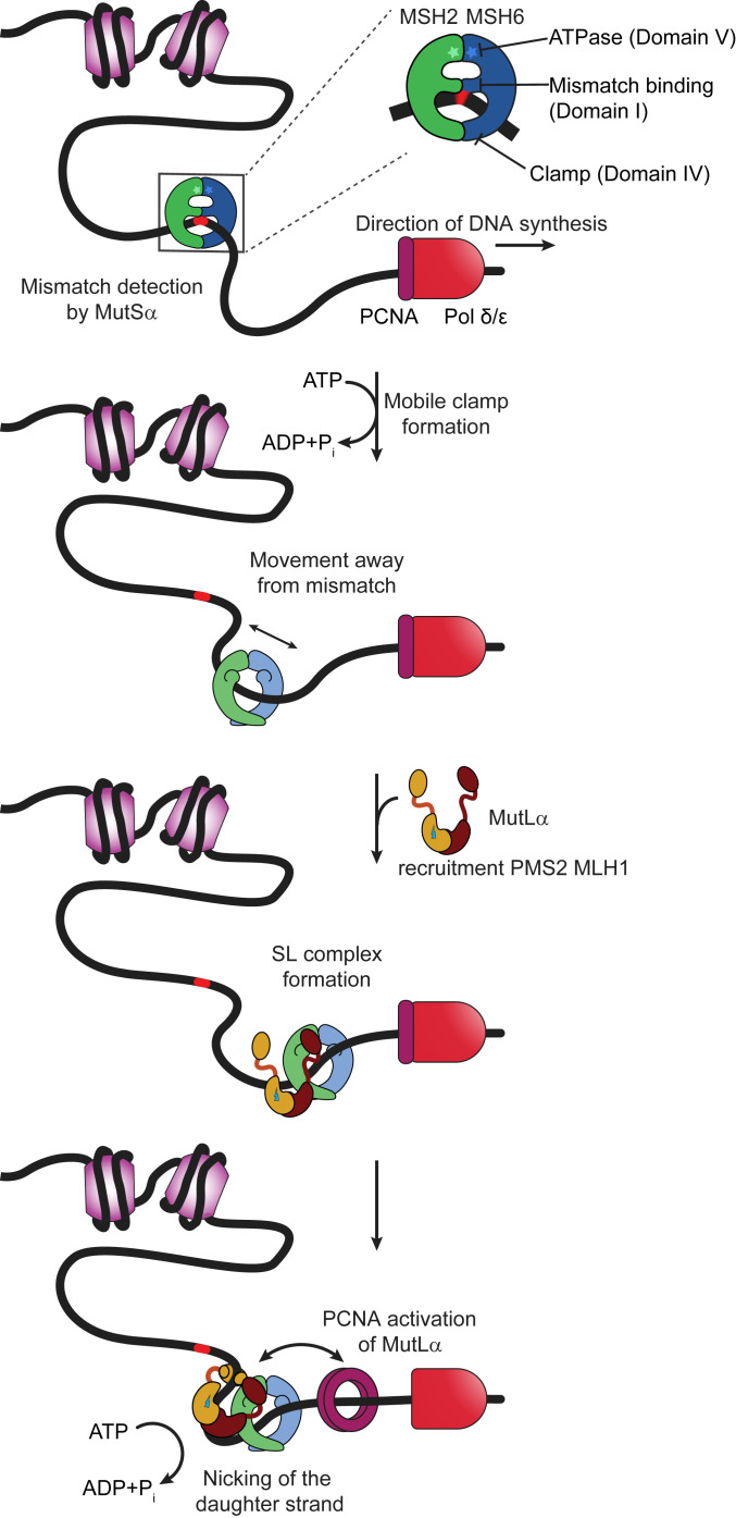
Fig. 1.
Schematic of mammalian mismatch repair initiation showing mismatch recognition, mobile clamp formation, MutLα recruitment, and PCNA activation of MutLα. Further description is provided in the text. MutSα is depicted as a green (MSH2) and blue (MSH6) theta-like dimer, with the ATPase sites indicated by stars at the top of the theta. The middle DNA binding domain of MSH6 interacts specifically with the mismatch. Nucleosomes are represented as light purple cylinders, DNA polymerase as red bullet, and PCNA as a dark purple ring. MutLα is depicted showing the C-terminal dimerization domains of MLH1 (burgundy) and PMS2 (ochre) connected by long flexible linker arms to the N-terminal DNA binding and ATPase domains. The endonuclease site on PMS2 is shown as a lightning bolt.

