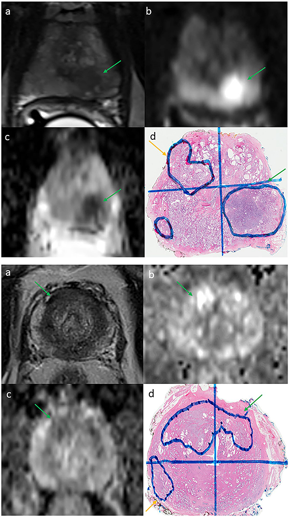
Figure 3.
(Top) Example of a missed discrete contralateral tumor in a 55-year-old man with a prostate-specific antigen (PSA) level of 7.5 ng/mL with a unilateral Prostate Imaging Reporting and Data System version 2 (PI-RADSv2) 5 target lesion (green arrow) with no evidence of a contralateral lesion on 3-tesla (3-T) multiparametric magnetic resonance imaging (mpMRI) or biopsy. (a) T2-weighted image. (b) Diffusion-weighted image. (c) Apparent diffusion coefficient map. (d) Detected (Gleason grade group 3 [GG3], green arrow) and missed (GG2, yellow arrow) contralateral prostate cancer lesions on final whole-mount histopathology slide. (Bottom) Example of a missed crossover tumor in a 54-year-old man with a PSA level of 5.3 ng/mL and a unilateral PI-RADS 3 target lesion (green arrow) with no evidence of contralateral extension on 3-T mpMRI. (a) T2-weighted image. (b) Diffusion-weighted image. (c) Apparent diffusion coefficient map. (d) Right-sided lesion with undetected contralateral extension (GG2, green arrow) and missed insignificant (GG1) prostate cancer on final whole-mount histopathology slide.

