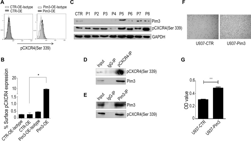
Figure 5.
PIM3 promotes the chemotaxis of acute myeloid leukemia (AML) cells via CXCR4. (A) Flow cytometry analysis of surface pCXCR4 (Ser339) expression on CTR-OE and Pim3-OE K562 AML cells. (B) Quantitative analysis of results from (A and C) Western blotting of PIM3 and pCXCR4 (Ser339) in samples from AML patients and healthy volunteers. (D) Immunoprecipitation of PIM3 and pCXCR4 (Ser339) and Western blotting of pCXCR4 (Ser339) and PIM3 (upper and lower panels, respectively). (E) Immunoprecipitation of PIM3 and pCXCR4 (Ser339) and Western blotting of PIM3 and pCXCR4 (Ser339) (upper and lower panels, respectively). (F) Imaging of control (U937-CTR, left panel) and PIM3-overexpressing (U937-Pim3, right panel) U937 cells subjected to a migration assay. (G) The cells in the lower migration chambers were collected and subjected to a quantitative CCK8 assay. *p< 0.05, ***p < 0.001.

