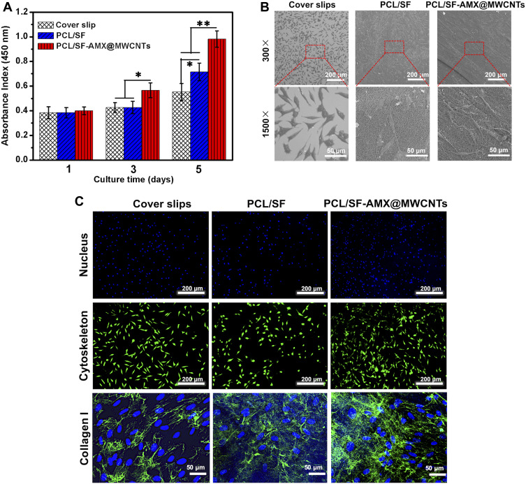
Figure 6.
The proliferation of L929 cells after culturing for 1, 3, and 5 days (A) and cell morphology on coverslips, PCL/SF mesh, and PCL/SF–AMX@MWCNT mesh after 3 days (B). FITC-conjugated phalloidin (green)/DAPI (blue) staining of cells, and collagen I (green) and DAPI (blue) staining of cells after culturing for 3 days on coverslips and various meshes accordingly (C). *P < 0.05 and **P < 0.01 in (A).

