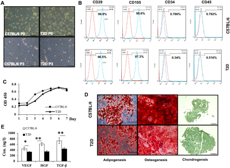
Fig. 3.
Characterization of ASCs isolated from C57BL/6 and T2D mice. a Representative micrographs of C57BL/6 and T2D ASCs at passage 0 and passage 3 observed under a light microscope. b Expression of CD29, CD105, CD34, and CD45 in ASCs harvested from C57BL/6L or T2D mice analyzed by flow cytometry. c Growth curves of C57BL/6 and T2D ASCs at passage 3. d The morphology of adipocytes, osteocytes, and chondrocytes derived from C57BL/6 ASCs and T2D ASCs identified by Oil Red, Alizarin Red, and Alcian Blue staining, respectively. Scale bar = 100 μm. e Concentrations of VEGF, HGF, and TGF-β secreted by C57BL/6 ASCs or T2D ASCs. Data are mean ± SEM of at least three individual experiments. At least 3 mice were included in each group. *P < 0.05, **P < 0.01, ANOVA test

