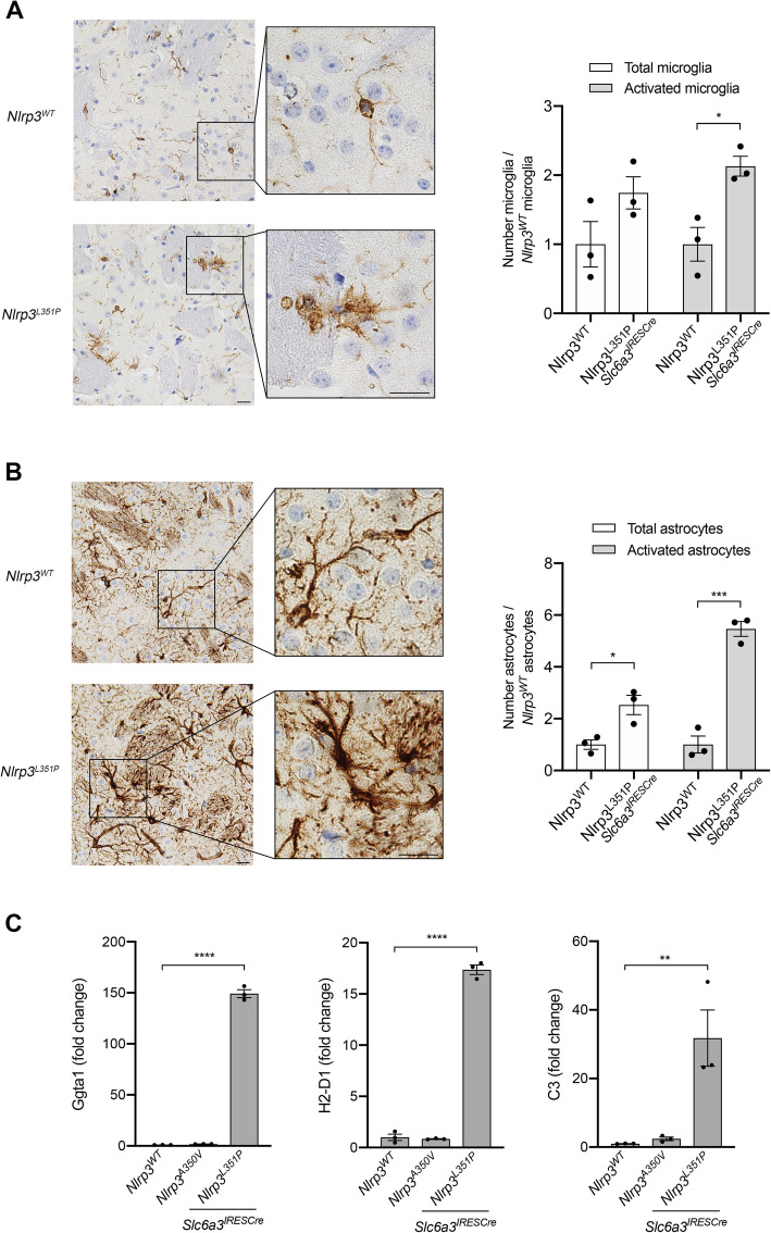
Fig. 6.
Evidence of enhanced proinflammatory glial cell phenotype in the striatum of Nlrp3L351P animals. a Striatal tissue sections from 18-month-old animals were probed with anti-IBA1 antibodies followed by BOND polymer detection. Microglial activation can be visualized in animals harboring the Nlrp3L351P allele (bottom panel), compared to their WT littermates (top panel). Scale bars represent 20 μM. There are significantly more activated microglia in tissues from animals expressing Nlrp3L351P compared with tissues from Nlrp3WT animals (* p = 0.0164, t test). Error bars represent s.e.m. b Adjacent striatal sections were stained with anti-GFAP antibodies. GFAP-immunoreactive astrocytes in Nlrp3L351P animals have an increased activation phenotype (bottom panel), compared to Nlrp3WT animals (top panel). Scale bars represent 20 μM. Quantitation revealed significantly more total astrocytes (* p = 0.0217, t test) as well as significantly more activated astrocytes (*** p = 0.0005, t test) in striatal tissues from Nlrp3L351P animals compared to controls. Error bars represent s.e.m. c Expression of A1 astrocyte markers, Ggta1, H2-D1, C3, were assessed in 18-month-old animals. RNA was isolated from striatal tissues, reversed transcribed, and mRNA was measured by real-time PCR. Ggta1, H2-D1, and C3 levels were significantly elevated in Nlrp3L351P mice compared to Nlrp3WT animals (**** p < 0.0001, **** p < 0.0001, ** p = 0.009 respectively, one-way ANOVA). Error bars represent s.e.m.

