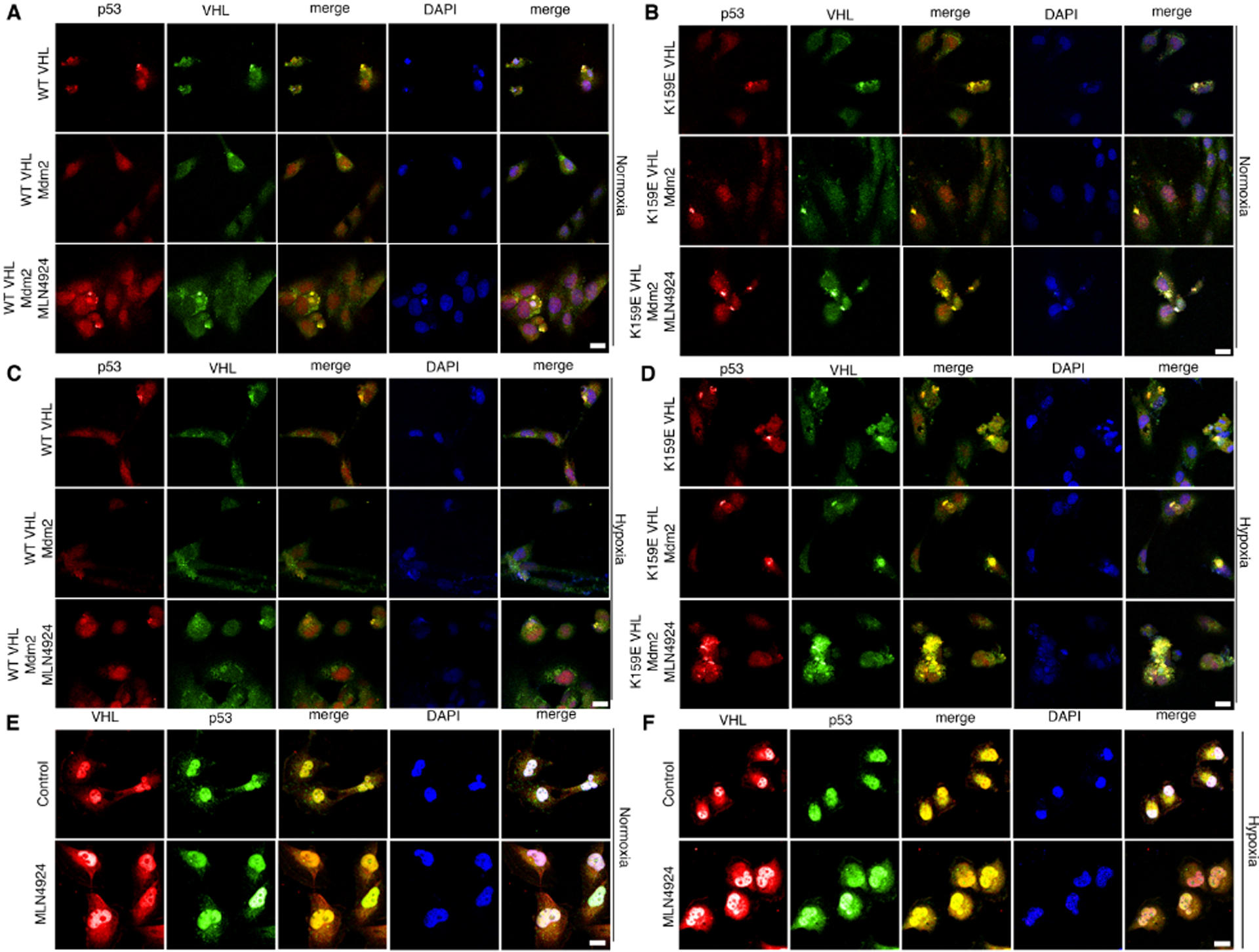
Figure 4. VHL forms a complex with p53 under hypoxia or with treatment of MLN4924.

(A-B) Transfection of 786-O cells with WT VHL (A) or K159E VHL (B) and Mdm2 under normoxia. Cells were treated with MLN4924 or a control, fixed, and stained with antibodies against p53 and VHL and subjected to immunofluorescence confocal microscopy. Scale bar represents 25 μm.
(C-D) Transfection of 786-O cells with WT VHL (C) or K159E (D) VHL and Mdm2 under hypoxia. Cells were treated with MLN4924 or a control, fixed, and stained with antibodies against p53 and VHL and subjected to immunofluorescence confocal microscopy. Scale bar represents 25 μm.
(E-F) Immunoflourescence microscopy of Caki-1 cells treated with MLN4924 or a control under normoxia (E) or hypoxia (F). Cells were fixed and stained with antibodies against VHL and p53. Scale bar represents 50 μm.
