
FIGURE 4.
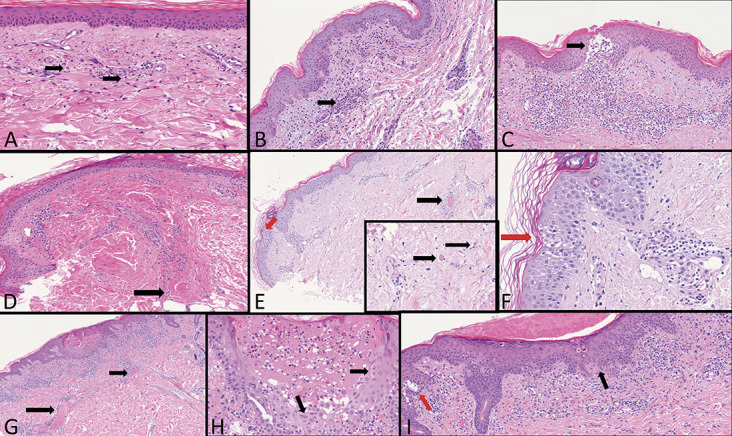
First group: COVID-19 active infection with PCR positive test: A, Highly dilated capillaries, initial blood extravasations, and some rare eosinophils (arrows). B, Dense perivascular lymphocytes cuffs around small capillary vessels (arrow). C, A large nest of Langerhans cells (arrow) in the superficial dermis dense perivascular lymphoid infiltration with extravasated red blood cells. D, A large thrombus in the deep vascular plexus (arrow). E, A large thrombus in the mid dermis (black arrow). Intraepidermal Langerhans cell nest (red arrow). Inset: Small thrombi in the deep dermis (arrows). F, Nest of Langerhans cells (red arrow). G, Extremely dilated vessels in the mid dermis in a Grover-like dermatosis (arrows). H, A vesicle-pustule surrounded by keratinocytes with cytopathic changes similar to herpetic infections (arrows). I, Sovra-epidermal clefts (red arrow), dyskeratotic keratinocytes, and patchy band-like infiltration with exocytosis (black arrow).