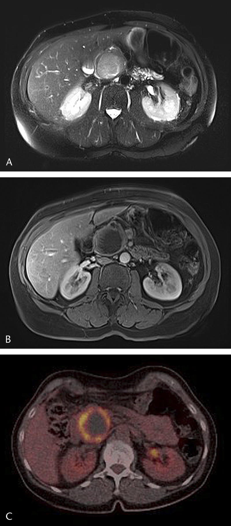
FIGURE 1.

Axial T2-weighted MR image (half-Fourier acquisition single-shot turbo spin echo [HASTE]) with a sharply demarked high signal intensity mass (45 mm) in the pancreatic head (A). Axial T1-weighted image (T1 volumetric interpolated breath-hold examination [T1-VIBE] with fat suppression) after intravenous injection of a contrast agent (gadoterate meglumine) shows enhancement of the wall with some linear papillary projections and a large nonenhancing center, possibly because of necrosis or mucinous component (B). On fluorine-18 fluorodeoxyglucose positron emission tomography (18F-FDG-PET), the wall of the tumor is avid with a large photopenic center (C).
