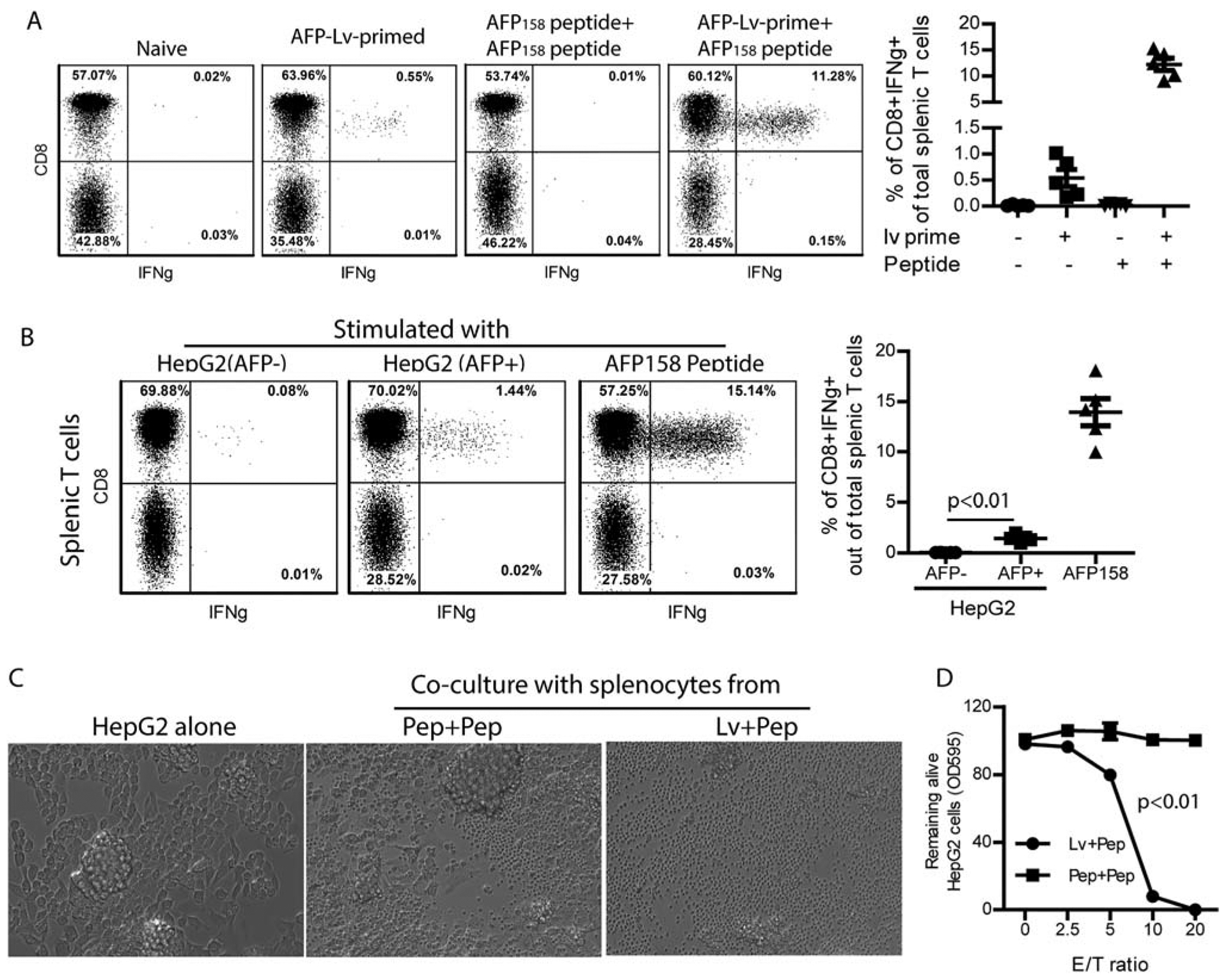
FIG. 1.

Immunization of AAD mice elicits a high level AFP158-specific CD8 T cells that recognize and kill human HepG2 cells. (A) HLA-A2 transgenic AAD mice were primed with AFP-lv and boosted with AFP158 peptide. Peripheral blood cells from the indicated mice were analyzed for CD8 and IFNγ 12 days after prime and 5 days after boost by gating on the Thy1.2+ T cells after ex vivo stimulation with AFP158 peptide. Representative dot plots and a summary of five mice are shown. (B) Splenocytes of the lv-primed and peptide-boosted mice were cocultured with AFP− or AFP+ HepG2 cells for 4 hours and analyzed for IFNγ. AFP158 peptide stimulation was used as positive control. Representative plots and a summary of five mice are shown. A t test was used for statistical analysis. (C,D) The cytotoxicity of the immunized mouse splenocytes was studied by coculturing them with HepG2 cells. Pictures were taken after overnight coculture (C). An MTT assay was then performed to determine the remaining live HepG2 cells after splenocytes were rinsed away. The MTT data of HepG2 tumor cells cocultured with different ratios of splenocytes was compared to MTT data of HepG2 tumor cells alone to calculate the percentage of live cells. Shown is the dose-dependent killing of HepG2 cells by splenocytes. Analysis of variance was used for statistical analysis. The experiment was done three times with similar data. Abbreviations: Lv+Pep, lv-primed and peptide-boost; Pep+Pep, peptide-prime and peptide-boost; OD, optical density.
