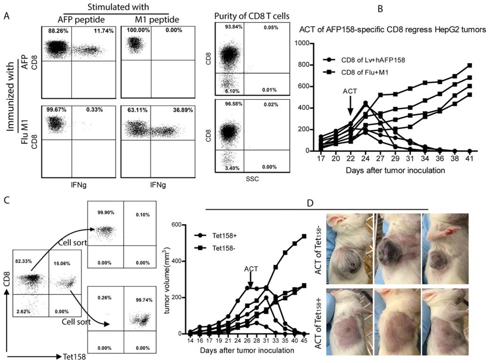
FIG. 3.

Adoptive transfer of AFP158-specific CD8 T cells eradicates HepG2 tumors in NSG mice. (A) CD8 T cells from AFP-immunized mice were isolated by magnetic beads. CD8 T cells from mice immunized with influenza virus M1 antigen peptide were used as control. The purity of CD8 T cells and the percentage of IFNγ-producing cells are shown. (B) NSG mice bearing HepG2 tumors were injected with 5 million CD8 T cells from AFP-immunized or M1-immunized mice (~0.5 million AFP-specific and 1.5 million M1-specific CD8 T cells, respectively). Tumor growth curve is shown. (C) Magnetic bead-purified CD8 T cells from AFP immunized mice were further separated into Tet158+ and Tet158− cells by cell sorting after Tet158 tetramer staining. The purity of Tet158+ and Tet158− CD8 cells before and after sorting is presented. (D) NSG mice bearing HepG2 tumors were injected with 0.5 million Tet158+ or Tet158− CD8 T cells. Tumor growth curve and pictures at the end of the experiment are presented. Abbreviations: ACT, Adoptive cell transfer; SSC, side scatter.
