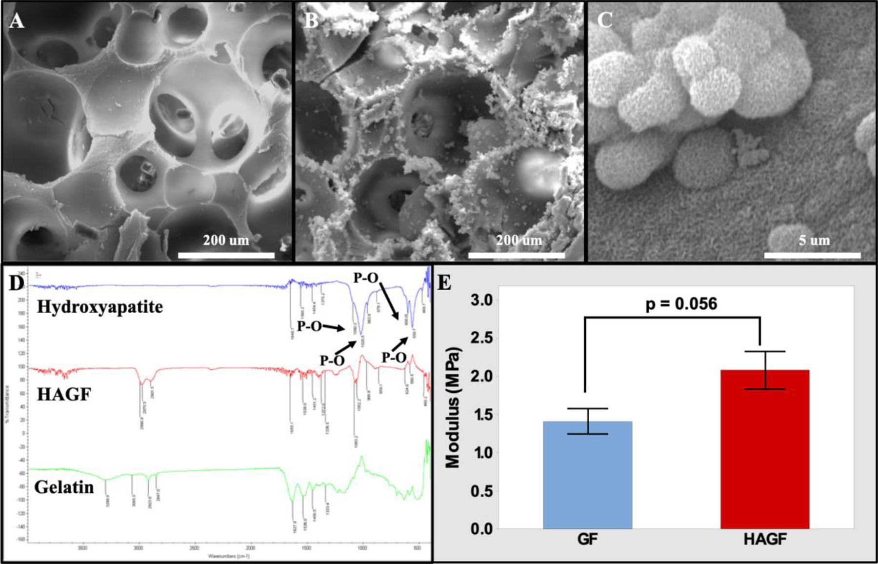
Figure 1.

Scanning electron microscopy Images of scaffolds. GF (A) and HAGF scaffold (B) at 500X and HAGF scaffold at 20000X (C) magnifications respectively. (D) FTIR peaks of gelatin type B (green), hydroxyapatite (blue), and HAGF scaffold (red). (E) HAGF scaffold showed stronger compression strength. HAGF: hydroxyapatite-coated nanofibrous gelatin.
