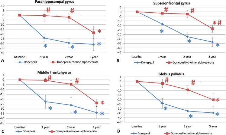
Fig. 6.
Changes in the parahippocampal gyrus (A), superior (B) and middle (C) frontal gyri, and globus pallidus (D) in the two groups of patients along the three years of observation. The data are the means of the percentage variation±S.E.M. *p < 0.05 versus baseline; #p < 0.05 versus monotherapy with donepezil alone.

