
FIGURE 1.
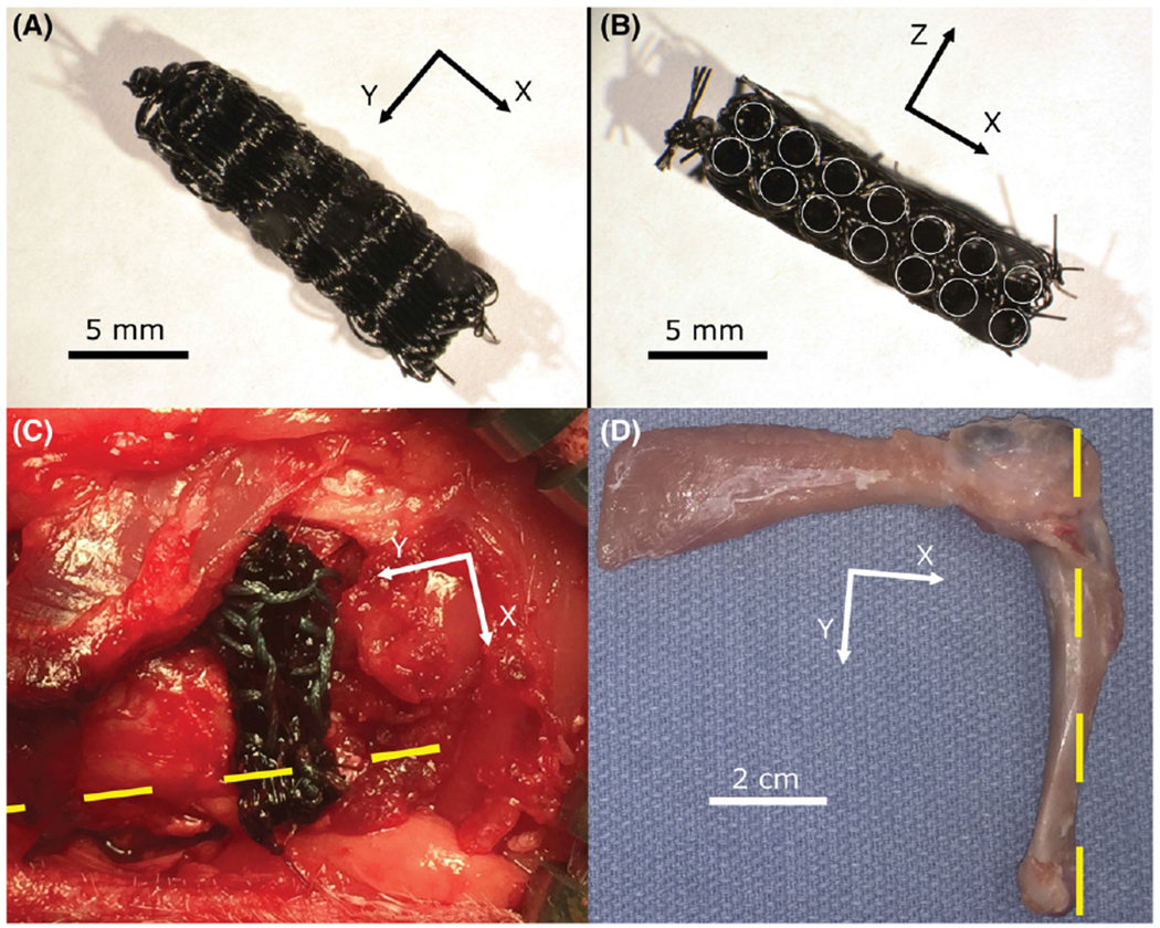
Scaffolds of electrochemically aligned collagen yarns were woven, stacked, and consolidated using a weft fiber to form a complete scaffold (A and B, circular holes highlighted in B). The complete, sterilized scaffold was inserted into a defect created within the right infraspinatus tendon of a rabbit and secured in place on the remaining tendon using 3–0 braided Ethibond suture in a Krackow pattern. The yellow dashed line highlights the longitudinal axis of the diaphysis (C). The humerus, scaffold, and infraspinatus tendon were harvested following 3 months of implantation (D).