
FIGURE 7.
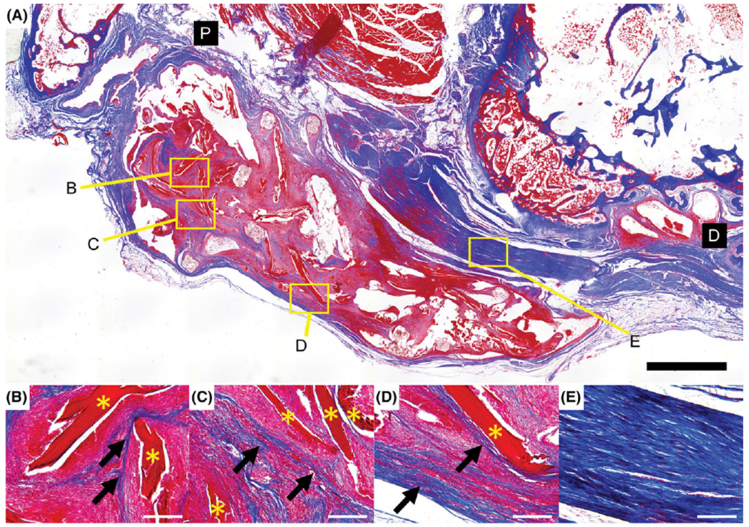
Specimen from cell-seeded scaffold group stained with Masson’s trichrome. Newly deposited collagen is visible within the continuum of the scaffold (arrows) following the contours of the ELAC fibers (*) (B–D). Extensive alignment of the collagen within the fibrous tissue surrounding the scaffold is also shown (E). Distal and proximal ends of the section are denoted by black boxes (D and P). Scale bar = 2 mm (A) and 200 μm (B–E).