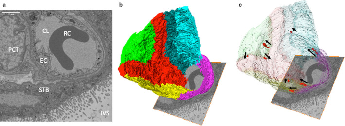
FIGURE 1.

Three‐dimensional reconstruction of a region of capillary from a terminal villus. (a) One of the 367 serial images in the SBF SEM stack used to reconstruct the capillary in three dimensions (12.6 × 12.6 × 18.35 μm). The image shows the capillary lumen (CL) containing a red blood cell (RC), the surrounding endothelial cells (EC), a pericyte (PCT), the syncytiotrophoblast (STB) and the intervillous space (IVS). Additional images from the stack can be seen in Appendix S1. (b) A three‐dimensional reconstruction of the stack showing the organisation of different endothelial cells, each of which is presented in a different colour. (c) The same image with the endothelial cells made transparent to reveal the location of IEPs (red) with their orientation highlighted by arrows pointing in the direction in which they protrude
