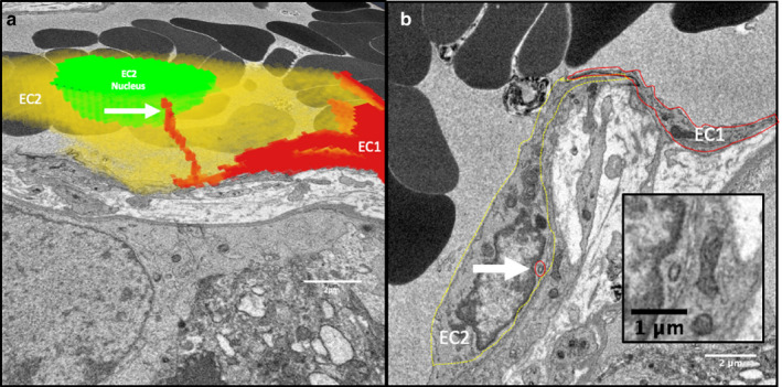
FIGURE 3.

IEPs extend from one endothelial cell into another. (a) A three‐dimensional reconstruction of two partial endothelial cells (red [EC1] and yellow [EC2]) based on 80 serial images 50 nm apart. An IEP from EC1 extends within EC2. (b) A single section from the image stack showing EC1 (solid red line) and EC2 (dashed yellow line) with the cross‐section of the IEP from EC1 within EC2 highlighted by a red circle. The inset shows a higher magnification image of the IEP cross‐section
