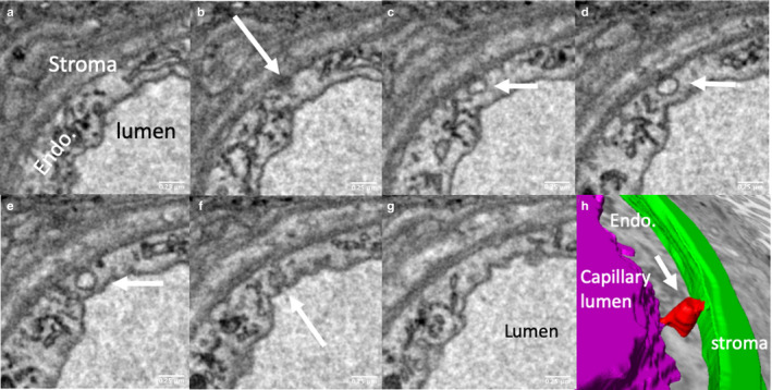
FIGURE 6.

Serial sections illustrating a putative trans‐endothelial channel. The trans‐endothelial channel indicated by white arrows in (b–f) opens on the abluminal side in (b) and on the luminal side in (f). (h) A three‐dimensional reconstruction of the channel (red), connecting the lumen (purple) and the villous stroma (green). The reticular structures indicated by black arrows in (c and g) appear to connect the luminal and abluminal surfaces; it is conceivable that these could be adapted to form channels
