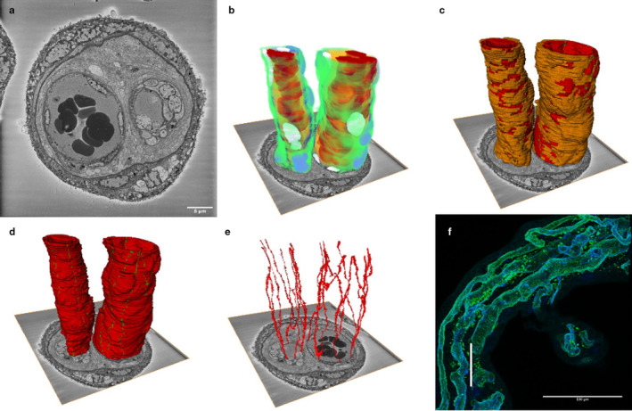
FIGURE 7.

Three‐dimensional reconstruction of an arteriole and venule from human term placenta. This image is based on 540 serial images (100 nm apart) from an arteriole and a venule pair from an intermediate villus. We suggest that the larger, lower resistance vessel on the right is the venule and the smaller vessel on the left the arteriole. (a) A representative image from the SBF SEM image stack. (b) A reconstruction of the arteriole and venule showing pericytes in green, pericyte nuclei in purple, endothelial cells in blue, endothelial nuclei in white, plasma in yellow and erythrocytes in red. (c) The arteriole and venule showing that pericytes (orange) cover a high proportion of both vessels. Endothelial cells are shown in red and the junctions in green. (d) The two vessels showing the endothelial cells in red and the junctions in green. (e) The endothelial cell junctions of the arteriole and venule. The difference in the arrangement of endothelial cells is most clearly seen when this image is rotated, and a movie is provided in Appendix S1. (f) To provide context and scale, this confocal image shows a similar region of intermediate villus with the vertical white scale bar representing the size of the SBF SEM stack (54 μm). The endothelium is blue and the pericytes green. There are three vessels in this villus; note the anastomosis between two of these vessels
