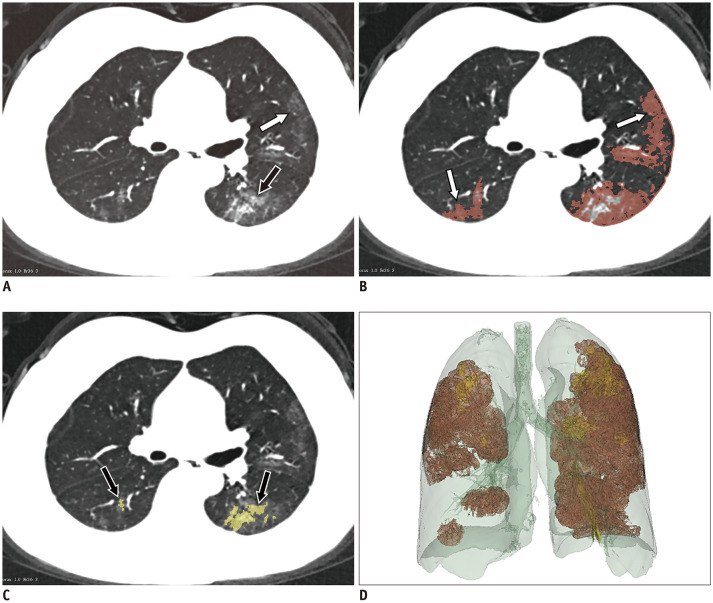
Fig. 3. GGOs and consolidation are segmented with 3D Slicer software on CT images.
A. GGO (white arrow) combined with consolidation (black arrow) in subpleural area. B, C. Manual delineation of scope of GGO (white arrows) and consolidation (black arrows) by threshold selection. D. Entire lesion fused with GGO and consolidation is shown in whole lung volume.

