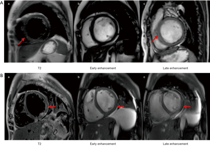
Figure S2.
CMR of the patients with PM and myocarditis. (A) is a 23-year-old female; (a) is sub-endocardial edema of the lateral and septal wall (arrow); (b) is no obvious early enhancement; (c) is diffuse sub-endocardial late enhancement of the septum, inferolateral and left ventricular free walls (arrow). (B) is a 43-year-old female; (a) is diffuse transmural edema of the left ventricle in T2-weighted image (arrow); (b) is early enhancement of the middle-layer myocardium in the inferolateral wall in T1-weighted image (arrow); (c) is diffuse late enhancement of the sub-endocardial and middle-layer myocardium in the septum, inferolateral and left ventricular free wall (arrow). CMR, cardiovascular magnetic resonance; PM, polymyositis.

