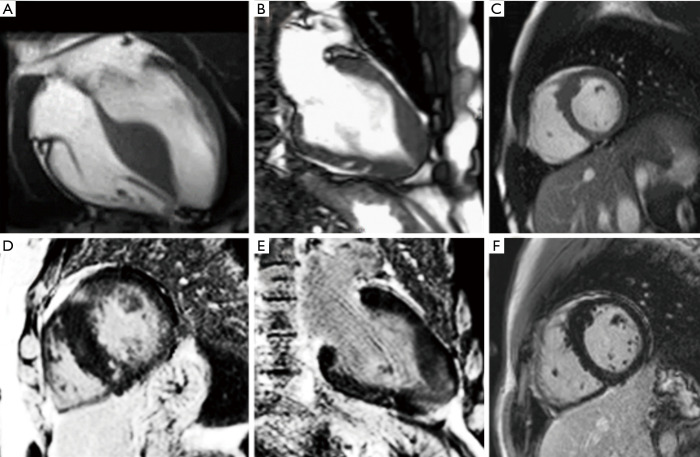
Figure 6.
The ability of CMR to show not only the different patterns of hypertrophic cardiomyopathy, (A) classical asymmetrical septal hypertrophy, (B) apical hypertrophy, (C) localised/discreet hypertrophy, but also the differing patterns of fibrosis (D) classical inferior and superior LV/RV junctions and region of hypertrophy, (E) apical fibrosis, (F) fibrosis in the hypertrophied section (in this case minor).

