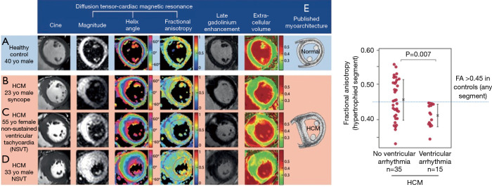
Figure 9.
Left panel: DTI-CMR can provide in vivo assessment of left ventricular myoarchitecture. The HA is the average myocyte orientation, and FA is a surrogate measure of underlying cell organization. The mid-ventricular slice at diastole in healthy control subjects (A) and patients with HCM (B,C,D) demonstrated similar HA distributions but marked differences in FA. These patterns were consistent with previously published HCM histology that shows disarray and fibrosis invading the mid-wall at the insertion point and hypertrophied segments (E); right panel: diastolic FA, measured in the hypertrophied segment, was reduced in HCM with ventricular arrhythmia compared with those without. Reproduced with permission and without changes under Creative Commons license from Ariga et al. (21). HA, helix angle; FA, fractional anisotropy.

