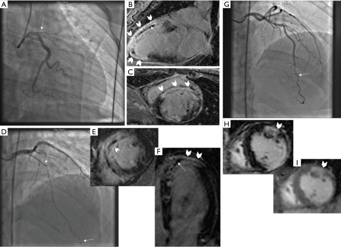
Figure 1.
CMR findings in patients with SCAD. Patient 1: (A) coronary angiogram showing ostial LAD SCAD involving large territory, (B,C) CMR findings of LGE (block arrows) and MVO (straight arrow). Patient 2: (D) Coronary angiogram showing diffuse mid-apical LAD SCAD (between straight arrows), (E,F) CMR findings of LGE (block arrows) and MVO (straight arrow). Patient 3: (G) Coronary angiogram showing distal LAD SCAD, (H) T1-weighted CMR showing LGE (block arrow), and (I) T2-weighted CMR showing myocardial edema (block arrow). CMR, cardiac magnetic resonance imaging; SCAD, spontaneous coronary artery dissection; MVO, microvascular obstruction; LAD, left anterior descending; LGE, late gadolinium enhancement.

