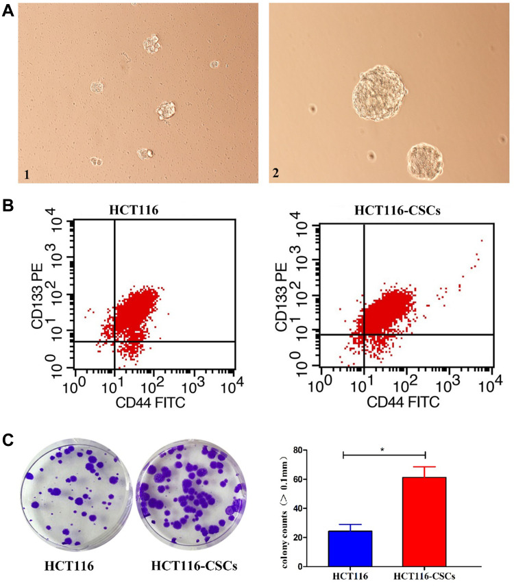
Figure 3.
Characterization and growth of HCT116-CSCs. (A) HCT116-CSCs culture (× 200). 1, HCT116-CSCs on day 5 after incubation; A2, HCT116-CSCs on day 5 after successive 6 passages of incubation; (B) Flow cytometry detects the percentages of CD133+CD44+ cells in parental HCT116 cells and HCT116-CSCs; (C) Colony-formation assay measures the colony formation of parental HCT116 cells and HCT116-CSCs. *P < 0.05.

