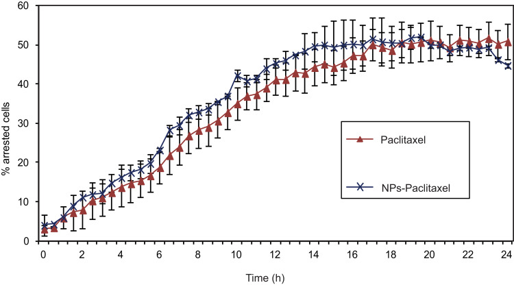
Figure 5.
The effect of Paclitaxel-loaded PPSu-PEG NPs on tubulin. HeLa K cells stably expressing GFP-α-tubulin/mcherry-histone H2B were imaged for 24 h with 10-minute intervals, after the addition of 250 nM free Paclitaxel or encapsulated in PPSu-PEG NPs, and the number of cell cycle arrested cells were counted. Graph demonstrates the % of arrested cells by either Paclitaxel or Paclitaxel-loaded PPSu-PEG NPs, as measured during 24 h of live cell monitoring. At least 100 cells were imaged, per condition per experiment.

