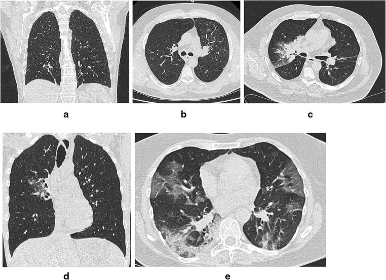
Fig. 1.
a CO-RADS 1: A few fibrotic bands in the lower lobes. No evidence of infection. RT-PCR−. b CO-RADS 2: Bronchial wall thickening, small centrilobular nodules, and tree in bud abnormalities in the left upper lobe. Consistent with bronchiolitis. RT-PCR−. c CO-RADS 3: Consolidation with surrounding ground glass opacity in right upper lobe. RT-PCR−. d CO-RADS 4: Bilateral areas of patchy ground glass opacity with associated small peribronchovascular consolidations. Predominantly central distribution. RT-PCR+. e CO-RADS 5: Bilateral peripheral ground glass abnormalities with areas of associated consolidation. RT-PCR+

