
Figure 8. Soft, deformable DexVS matrices promote MF induction.
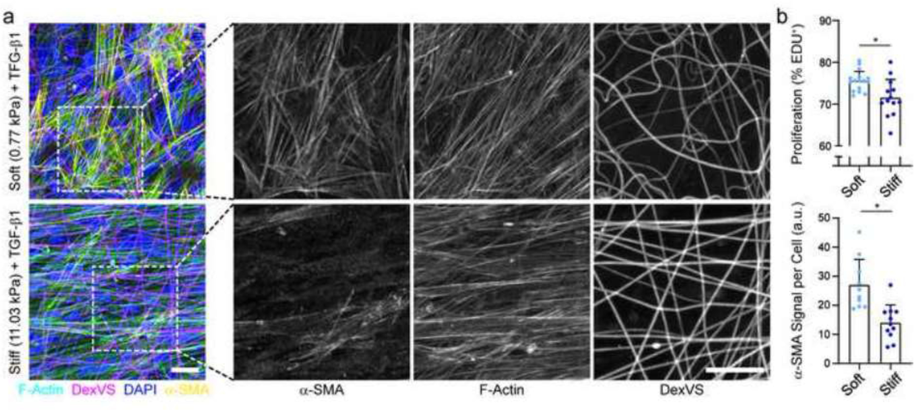
(a) Confocal fluorescent image of NHLFs cultured for 7 days on soft and stiff DexVS fibrous matrices; F-actin (cyan), DexVS fibers (magenta), nuclei (blue), α-SMA (yellow). Dashed boxes indicate locations of higher magnification images depicting α-SMA stress fibers. (b) Quantification of cell proliferation by EdU labeling (n ≥ 13) and α-SMA fluorescent intensity (n ≥ 10). Scale bars: 25 μm. All data presented as mean ± std; * p ≤ 0.05.