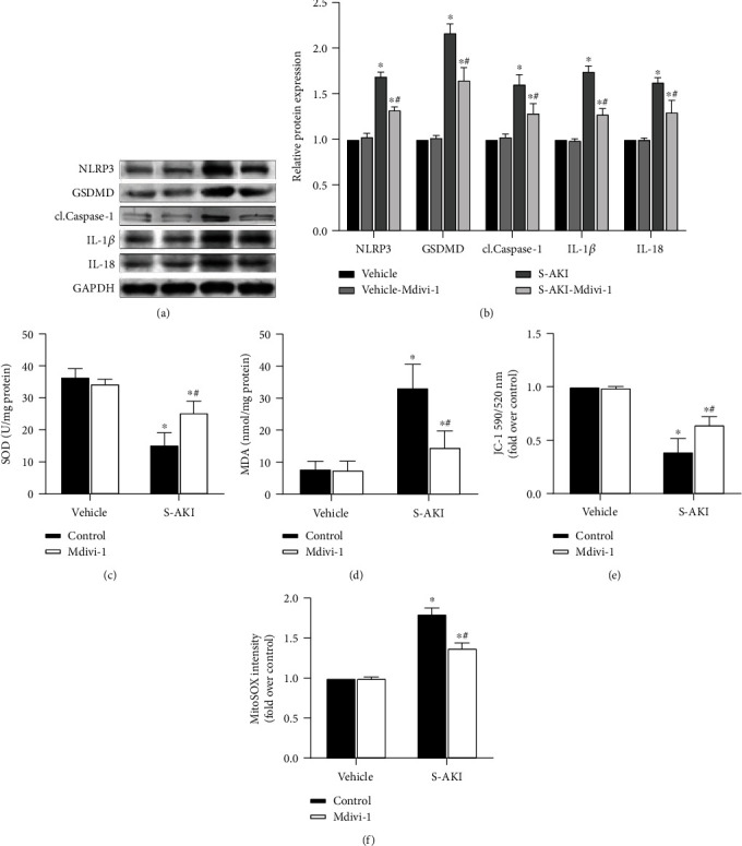
Figure 2.

S-AKI mouse model shows that inhibition of DRP1 expression attenuates oxidative damage to kidney tissue and protects mitochondrial function by NLRP3 inflammasome pathway protein expression downregulation. (a) Western blot protein expression bands of the NLRP3 inflammasome-related proteins, NLRP3, GSDMD, cl.Caspase-1, IL-1β, and IL-18. (b) Semiquantitative analysis of Western blot protein expression bands of the NLRP3 inflammasome-related proteins. (c) Comparison of superoxide dismutase (SOD) levels in renal tissue homogenate between different treatment groups. (d) Comparison of malondialdehyde (MDA) levels in renal tissue homogenate between different treatment groups. (e) JC-1 staining of the purified mitochondria, which represents the mitochondrial membrane potential level in the different groups. (f) ROS staining of the purified mitochondria, which represents the reactive oxygen species level in mitochondria in the different groups. n = 6 mice in each group for all experiments. The data are presented as means ± SEM. ∗P < 0.05 versus control-treated mouse group. #P < 0.05 versus LPS-induced S-AKI group.
