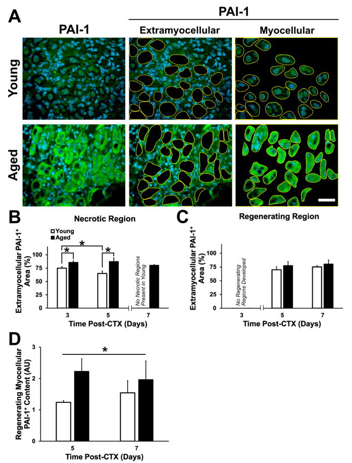
Figure 9.
PAI-1 expression is greater in aged skeletal muscle. (A) Immunostaining of PAI-1 (green) and DAPI (blue) at five days following damage. (B) A significant interaction between age and recovery time point following damage was observed in the necrotic regions (p = 0.044). Simple main effect post-hoc analyses demonstrated a significant greater extramyocellular PAI-1 expression within aged muscle at three and five days following damage (p = 0.005 and p < 0.001, respectively). Extramyocellular PAI-1 within the necrotic regions of young muscle declined significantly between three and five days following damage (p = 0.015). * denotes significant differences between groups identified by the simple main effects post-hoc analyses (p < 0.05). (C) No significant differences in extramyocellular PAI-1 were observed in the regenerating region of young and aged muscle (p > 0.05). (D) Brightness analysis of PAI-1 within regenerating myofibers showed significantly greater PAI-1 signal within aged regenerating myofibers compared to young (* denotes main effect of age: p = 0.029). n = 4–5 per group. Data presented are means ± standard deviation. Scale bar represents 50 μm.

