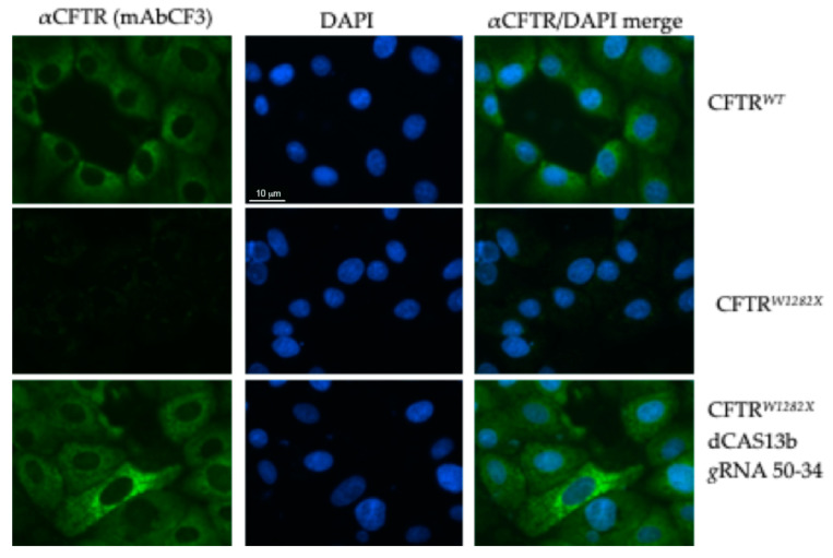
Figure 6.
Immunofluorescence detection of CFTR protein in FRT-CFTRW1282X cells. Cells untransfected (negative control) and transfected with the plasmids encoding the indicated gRNA and Cas13b/ADAR2DD (300 ng total DNA) are shown. FRT-CFTRWT cells were used as a positive control. Cells were fixed with methanol and the CFTR protein was revealed by the primary antibody mAbCF3, followed by a secondary antibody anti-mouse Alexa-488, (green, Abcam). Nuclei (blue) were DAPI stained. Images were taken at 100x magnification on a ZEISS microscope equipped for epifluorescence.

