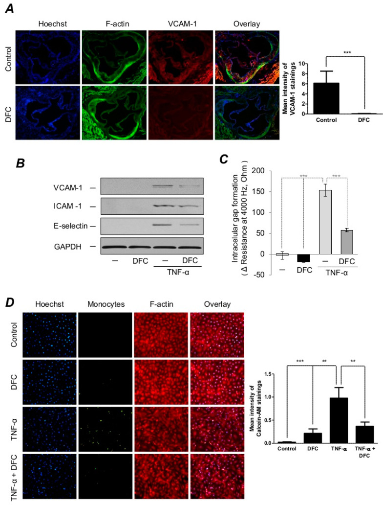
Figure 3.
DFC prevents TNF-α-induced expression of adhesion molecules in ApoE−/− mice and endothelial cells, preserves endothelial integrity and inhibits monocyte adhesion. (A) 8-week-old ApoE−/− mice were fed ad libitum with an atherogenic diet containing 5% fat and 1.25% cholesterol and divided into DFC group (n = 17) receiving 160 mg/kg intraperitoneal DFC and control group (n = 21) receiving physiological saline every second day for 8 weeks. Aortas (5 per group) were stained for DNA (Hoechst 33258, blue), F-actin (cytoskeleton, phalloidin-FITC, green) and VCAM-1 (AF647, red), respectively. Quantification of VCAM-1 mean intensity was performed with Image J software. Scale bar: 0.2 mm. (B) Human Umbilical Vein Endothelial Cells (HUVEC) were incubated with or without DFC (50 µmol/L) for 16 h and treated with or without 1 ng/mL of TNF-α for 8 h. Representative Western blots showing VCAM-1, ICAM-1 and E-selectin protein expression in HUVEC normalized to GAPDH are shown. (C) HUVEC were cultured with or without DFC (50 µmol/L) for 16 h in CM199 medium containing 5% FBS. Then, cells were incubated in the presence or absence of TNF-α (1 ng/mL) for 3 h. Transendothelial electrical resistance was monitored by the Electric Cell-substrate Impedance Sensing (ECIS) Zθ instrument over 3 h. (D) Confluent HUVEC were incubated with or without DFC (50 µmol/L) for 16 h in CM199 medium containing 5% FBS. Then, cells were incubated with or without TNF-α (1 ng/mL) for 6 h followed by coincubation with calcein-labeled (5 µmol/L) monocytes for 30 min at 37 °C. Cells were then fixed and stained for DNA (Hoechst 33258, blue) and F-actin (cytoskeleton, red, iFlour 647). Quantification of Calcein-AM mean intensity was performed with Image J software. Images were taken with a fluorescent microscope at a magnification of 400×. ** p < 0.01; *** p < 0.001.

