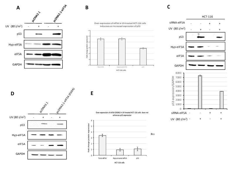
Figure 3.
eIF5A regulates p53 protein expression. (A) HCT-116 cells were transiently transfected with the pcDNA 3.1- eIF5A expression vector and the control plasmid pcDNA3.1, respectively. Twenty-four hours after transfection, cells were UV-C irradiated and were then grown for another 24 h. Cell lysates were analyzed by immunoblotting with the indicated antibodies. The expression level was normalized to GAPDH. (B) The intensity of bands was analyzed by the Bio-rad Image Lab Software 5.2.1 and the values of the histogram represent the fold change expression of indicated proteins in UV treated cells comparing their expression in HCT-116 cells transfected with the specific constructs with respect the empty one. Data are means ± SD (n = 3) (C) HCT-116 cells were transfected with siRNA targeting eIF5A mRNA 24 h before UV-C irradiation and were then grown for another 24 h. Cell lysates were analyzed by immunoblotting with antibodies to p53, hypusinated form of eIF5A and total eIF5A. Histogram represents p53 expression level normalized to GAPDH. (D) Over-expression of the construct pcDNA3.1-eIF5A(K50A) containing single-point mutation and UV-C irradiation of the cells. Cell lysates were analyzed by immunoblotting with antibodies to p53, hypusinated form of eIF5A and total eIF5A. The expression level was normalized to GAPDH. (E) The intensity of bands was analyzed by the Bio-rad Image Lab Software 5.2.1 and the values of the histogram represent the fold change expression of indicated proteins in UV treated cells comparing their expression in HCT-116 cells transfected with the specific constructs with respect the empty one. Data are means ± SD (n = 3).

