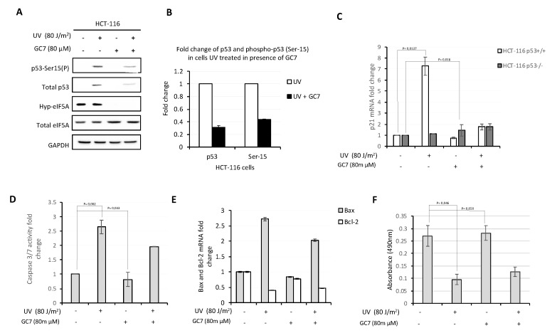
Figure 6.
GC7 counteracts pro-apoptotic effects of p53. (A) Analysis of p53 phosphorylation levels on Ser15 in HCT-116 cells treated with UV and/or GC7 performed by immunoblotting with the indicated antibodies. (B) Intensity of bands was detected by the Bio-rad Image Lab Software 5.2.1 and the signal of interest was normalized to control GAPDH. The values of p53 and p53-Ser15(P) in untreated cells was set as 1 and correspond to the control. The intensity of the signals in treated cells was calculated versus control group and reported as their ratio. Data are means ± SD (n = 4). (C) qPCR analysis of p21 mRNA expression in HCT-116 and HCT-116 p53−/− cells after UV and GC7 treatment. Results are presented in terms of a fold change after normalizing with GAPDH mRNA. Each value represents the mean ± S.D. of three independent experiments. p values reported to the upper side of the histograms were calculated versus control group. (D) Caspase 3/7 activity was measured in HCT-116 cells after UV and/or GC7 treatment as indicated. Each value represents the mean ± S.D. of three independent experiments. The value of Caspase 3/7 activity in untreated cells was set as 1. p values reported to the upper side of the histograms were calculated versus control group. (E) qPCR analysis of Bax and Bcl-2 mRNA in HCT-116 cells treated as indicated. Each value represents the mean ± S.D. of three independent experiments. Results are presented in terms of a fold change after normalizing with GAPDH mRNA. The value of target mRNAs in untreated cells was set as 1. (F) The viability of HCT-116 cells after UV and/or GC7 treatment was evaluated via MTS assay. Each value represents the mean ± S.D. of three independent experiments and p values reported to the upper side of the histograms were calculated versus control group.

