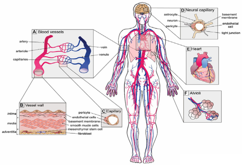
Figure 1.
Unique vascular beds in the human body. (A) Blood vessels are zonated and display unique cellular phenotypes and functionality. The 5 major zonation states of vessels are arteries, arterioles, capillaries, venules and veins. (B) Walls of arterial vessels are typically composed of 3 layers: tunica intima, tunica media and tunica adventitia. The intima is the innermost layer formed by endothelial cells that are in direct contact with the blood. The intima layer is mounted on the basement membrane, which is filled with fibro-elastic extracellular matrix, pericytes and smooth muscle cells. Media, the middle contractile layer, is composed of smooth muscle cells that provide support and flexibility to the vessel. Adventitia, the outmost layer of connective tissue surrounding the vessel, contains fibroblasts, a few mesenchymal stem cells and neurons. (C) Capillaries, the smallest blood vessels, are involved in direct solute exchange with the tissue. Capillaries possess a single layer of ECs that is surrounded by basement membrane and contains extracellular matrix and pericytes. Pericytes regulate the permeability of capillaries and their precise density varies from organ to organ. (D) Neural capillaries are characterized by an unfenestrated structure and ECs with tight junctions. Neural capillaries are densely populated by pericytes and are often contacted by astrocytes and microglia. (E) The heart is the central organ in the cardiovascular system that pumps blood through the whole body, and its function is supported by coronary arteries. (F) Lungs possess specialized vasculature that enables oxygen and carbon dioxide exchange between alveoli and pulmonary capillaries.

