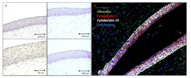
Figure 2.
Human skin equivalent generated using PCS-200-010 and PCS-201-010 cell lines and differentiated for 14 days. Sections of the formalin-fixed, and paraffin-embedded, 3-dimensional (3D) skin models. (A) Hematoxylin and Eosin staining. HSEs consist of well-differentiated epidermis on top of a fibroblast-populated dermis. (B) Fluorescent immunostaining. Green—Vimentin (Biolegend 677807), Red—Cytokeratin 1 (LSBio LS-C180221), White—Cytokeratin 14 (Abcam ab77684), Blue—Cell nucleus (DAPI). Cytokeratin 14 is strongly expressed in keratinocytes forming a basal layer with lower expression in the keratinocytes of the more apical layers, stratum spinosum, and stratum granulosum. Cytokeratin 1 expression is detected in the fully differentiated epidermis.

