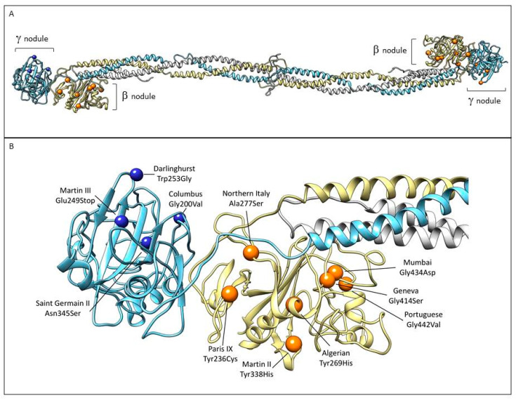
Figure 5.
Localization of the mutations associated with thrombosis within the beta and gamma nodules in the fibrinogen structure. (A) The three chains are colored as in Figure 1. Mutations associated with thrombotic complications in β nodule (orange sphere) and γ nodule (blue spheres) are indicated (Table 2). (B) Close-up view of the β and γ nodules and the described mutations. Images were produced using UCSF Chimera package (http://www.rbvi.ucsf.edu/chimera) [73] and the 3GHG coordinates.

