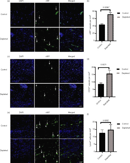
Figure 7.

Effect of depletion of langerin+ cells on blood and lymphatic vessels in the granulation tissue. The day 6 tissues were stained with vessel marker [von Willebrand factor (vWF), CD31 or Lyve‐1 – green] and nuclear stain DAPI (blue). Representative images of (a) vWF+, (c) CD31+ and (E) Lyve‐1+ vessels (not the cells) in the granulation tissue of control and depleted groups at day 6 post‐wounding (n = 8 wounds). The scale bar represents 50 μm. The total numbers of (b) vWF+, (d) CD31+ and (f) Lyve‐1+ per mm2 were quantified and presented as mean ± SEM (n = 8 wounds). Significant differences between the groups determined by Mann–Whitney U‐test are indicated with an asterisk (*P < 0·05).
